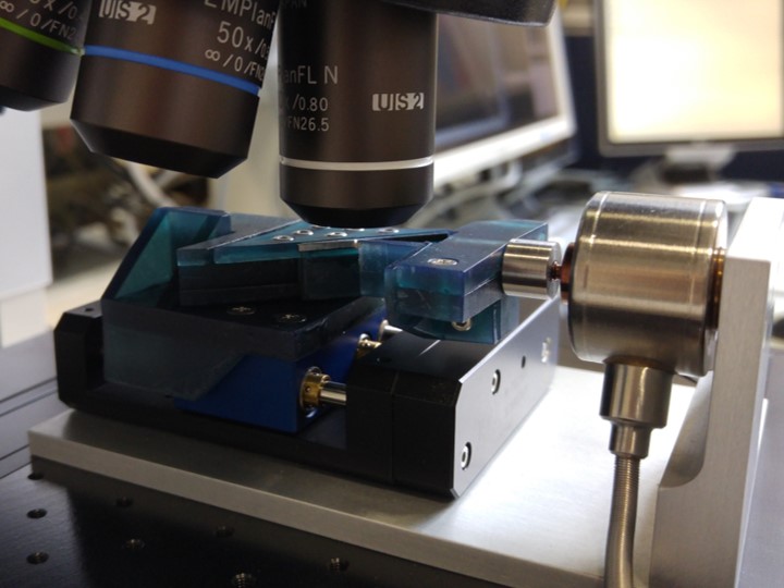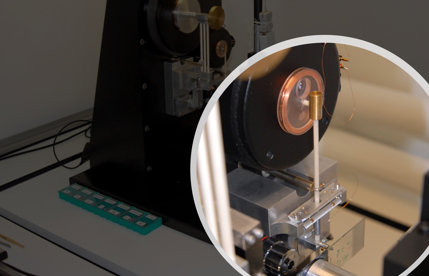


Laboratory mechanical tests on small size specimens:
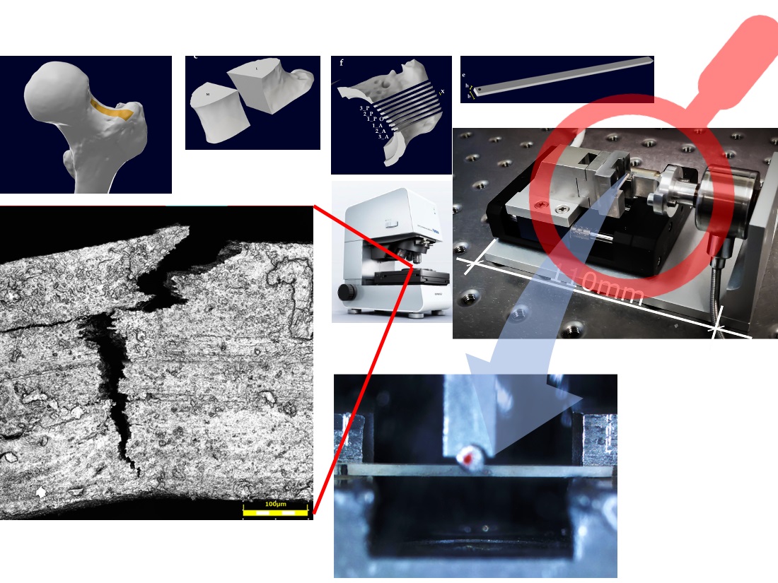
Surface analyses:
In-situ testing and Digital Image Correlation (DIC)
Numerical modeling complementing micromechanical testing: finite element modeling, homogenization approaches for mechanical and physical properties, numerical simulation of laboratory mechanical tests to determine deformation mechanisms at the small scale, micro-Computer-Tomography based finite element modeling, multigrid codes for computational micromechanics on micro-CT based models, multi-physics modeling.
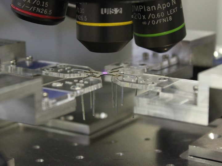
Development of new in-situ experimental techniques at sub-millimetre length scale, integration of mechanical tests and imaging (optical microscopy and confocal laser scanning microscopy).
The application contexts are the micromechanics of tissues and (bio)materials: relationship between structure and property. Assessment of the mechanical behaviour of materials and tissues on the basis of the micro-scale composition and of the relevant deformation mechanisms of the single constituents. Application of the micromechanics concepts and methods to biological tissues and (bio)materials.
As representative examples, tissues of the musculoskeletal system are characterized such as articular cartilage and bone tissue. Among the biomaterials, representative examples are: glass ceramic scaffolds, polyurethane scaffolds, micro- and nano-silk based vascular grafts, bovine pericardial tissue for heath valves, materials and structures obtained through Additive Manufacturing techniques.
Some of the laboratory facilities belong to the inter-department laboratory (IS-MicroLab) in which other industrial applications can be considered, es: MEMS.
ILARIA GUIDETTI
DEEPTHISHRE GUNASHEKAR
ILARIA ROTA